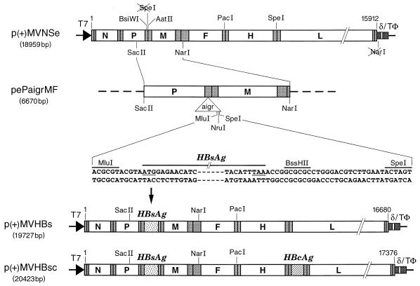
FIG. 1.
Cloning HBsAg and HBcAg ORFs in p(+)MVNSe antigenomic MV plasmid. ORFs of MV and HBV are shown as rectangles (not to scale) labeled with letters as follows: N, nucleocapsid; P, phosphoprotein; M, matrix; F, fusion; H, hemagglutinin; and L, large protein of MV. Stippled rectangles denote nontranslated regions, and vertical bars denote the nontranscribed intergenic trinucleotides. The triangle labeled “aigr” represents the artificial intergenic region, which consists of gene termination, intergenic, and gene start sequences followed by unique cloning sites. The flanking sequences together with start and stop codons (underlined) of the HBsAg ORF, plasmid names together with total sizes in base pairs, MV antigenomic nucleotide numbers (based on EMBL accession no. Z66517), and restriction sites are as indicated. T7 indicates the T7 RNA polymerase promoter, δ indicates the hepatitis delta virus ribozyme, and TΦ indicates the T7 RNA polymerase terminator.

