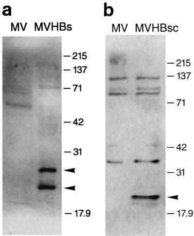
FIG. 3.
Western immunoblots of HBsAg and HBcAg expressed from recombinant MVs. Vero cells were infected with either MVHBs, MVHBsc, or MV-tag-Edm (indicated as MV), and total cellular proteins were harvested at 24 hpi and processed for Western blots as described in Materials and Methods. The nylon membranes with transferred proteins were developed with goat anti-HBsAg antibodies followed by anti-goat antibody–HRPO conjugate (a) and with rabbit anti-HBcAg antibodies followed by anti-rabbit antibody–HRPO conjugate (b), and proteins were visualized by enhanced chemiluminescence. Two protein bands of approximately 27 and 24 kDa observed in MVHBs cell lysates and a single band of approximately 21 kDa observed in MVHBsc cell lysates are marked with arrowheads. The molecular mass markers (in kilodaltons) are indicated.

