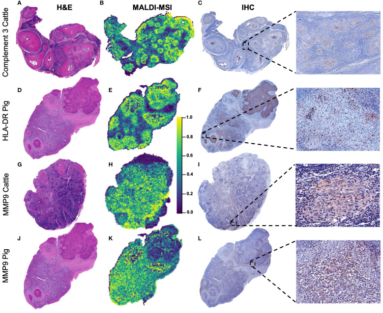
Figure 7.
Spatial distribution according to selected m/z features in bovine and porcine granulomas and validation by immunohistochemistry. (A, D, G, J) Sub-gross H&E-stained sections from a lymph node with multiple granulomas belonging to cattle or pigs, as specified. (B, E, H, K) Distribution pattern of C3 (m/z = 2,217.25) in a bovine lymph node (LN), HLA-DR (m/z = 1,963.90) in a porcine LN, and metalloproteinase 9 (MMP9) (m/z = 959.40) in bovine and porcine LNs. C3 and HLA-DR were homogeneously distributed throughout granulomas, whereas MMP9 was absent in the necrotic center of granulomas. (C, F, I, L) Immunolabeling against the three selected molecules in the different granulomas in each species to confirm matrix-assisted laser desorption/ionization mass spectrometry imaging (MALDI-MSI) results.

