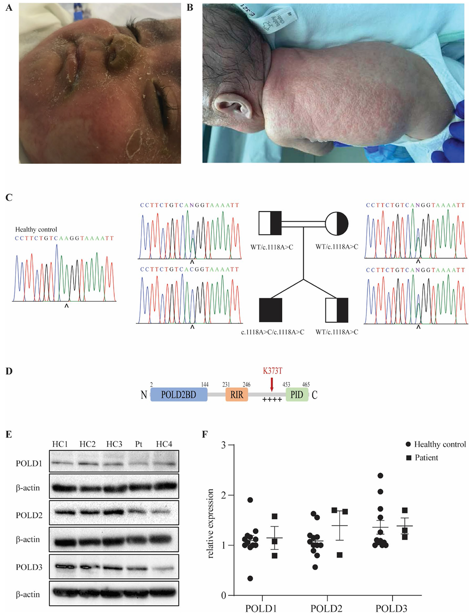
Fig. 1.

Identification of a homozygous mutation in POLD3 in a patient presenting with severe combined immunodeficiency and developmental defects. A Skin eczema due to neutrophil infiltration in the patient. B Skin infection with pustulosis and clinical alopecia in the patient. C Patient pedigree and familial segregation of the identified POLD3 mutation by Sanger sequencing compared to a healthy control. D Schematic representation of POLD3 domain structure. POLD2BD, POLD2 binding domain; RIR, RIV-1 interacting region; + + + + , disordered positively charged region; PID, PCNA interacting domain. The identified K373T mutation site is indicated in red. E Protein expression levels of POLD1, POLD2, and POLD3 in primary fibroblasts from four healthy controls (HC) and the patient (PT). β-actin was used as a loading control. The blot is representative of 3 independent experiments. F Quantification of POLD1, POLD2, and POLD3 protein expression in primary fibroblasts of four healthy controls and the patient normalized with β-actin expression (n = 3). Results represent mean ± SEM
