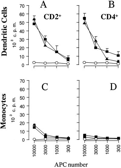
FIG. 3.
DC sensitize BHV-1-specific T lymphocytes in vitro. Increasing numbers of DC or monocytes, incubated with BHV-1 LAM, were cultured with 2 × 10⁵ autologous CD2+ (A and C) or CD4+ (B and D) lymphocytes. Proliferation was assessed by thymidine incorporation during the last 10 h of 5 days of culture. The data are expressed as counts per minute (c.p.m.), and each point represents the mean ± standard deviation of triplicate cultures. The results are representative of three independent experiments. Open circles, mock-infected APC; closed triangles, APC incubated with live BHV-1; closed squares, APC incubated with UV-inactivated BHV-1.

