Fig. 6.
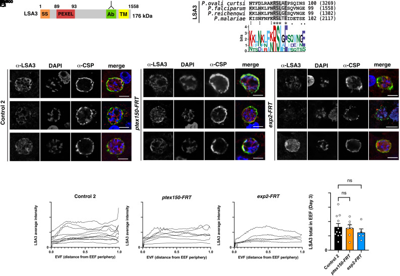
Localization of PEXEL protein LSA3 in P. falciparum EEFs. (A) Schematic of P. falciparum LSA3 including SS, PEXEL, Ab region, TM domain. (B) Multiple sequence alignment of LSA3 from Plasmodium species shows the PEXEL motif is conserved. (C) Immunofluorescence microscopy of LSA3 in P. falciparum Control 2, ptex150-FRT, and exp2-FRT EEFs on day 3 postinfection. (Scale bar, 5 mm.) (D) Quantification of LSA3 distance from the parasite periphery in P. falciparum Control 2, ptex150-FRT, and exp2-FRT EEFs on day 3 postinfection. Data are mean ± SEM from n = 5 to 11 individual EEFs per condition analyzed by one-way ANOVA (Kruskal–Wallis test). P values are shown; ns, not significant.

