
FIGURE 1.
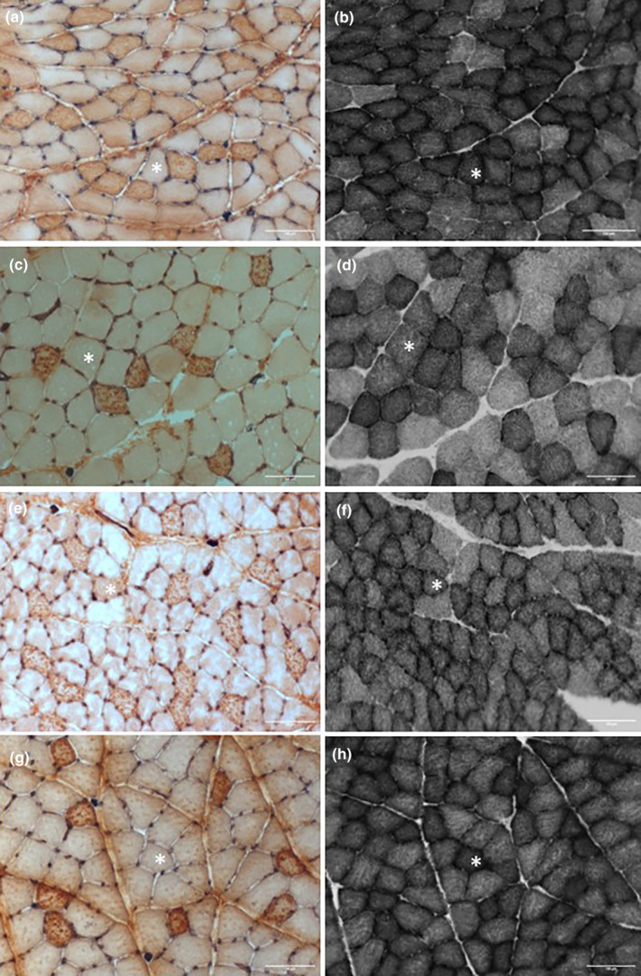
Histological pictures of serial sections from the m. extensor carpi radialis (ECR) (a, b, e, f) and m. flexor carpi ulnaris (FCU) (c, d, g, h) of control (a–d) and stroke (e–h) rats stained for capillaries and type I (dark) fibers (a, c, e, g), and succinate dehydrogenase (b, d, f, h). *Same fiber in serial section; scale bars 100 μm.