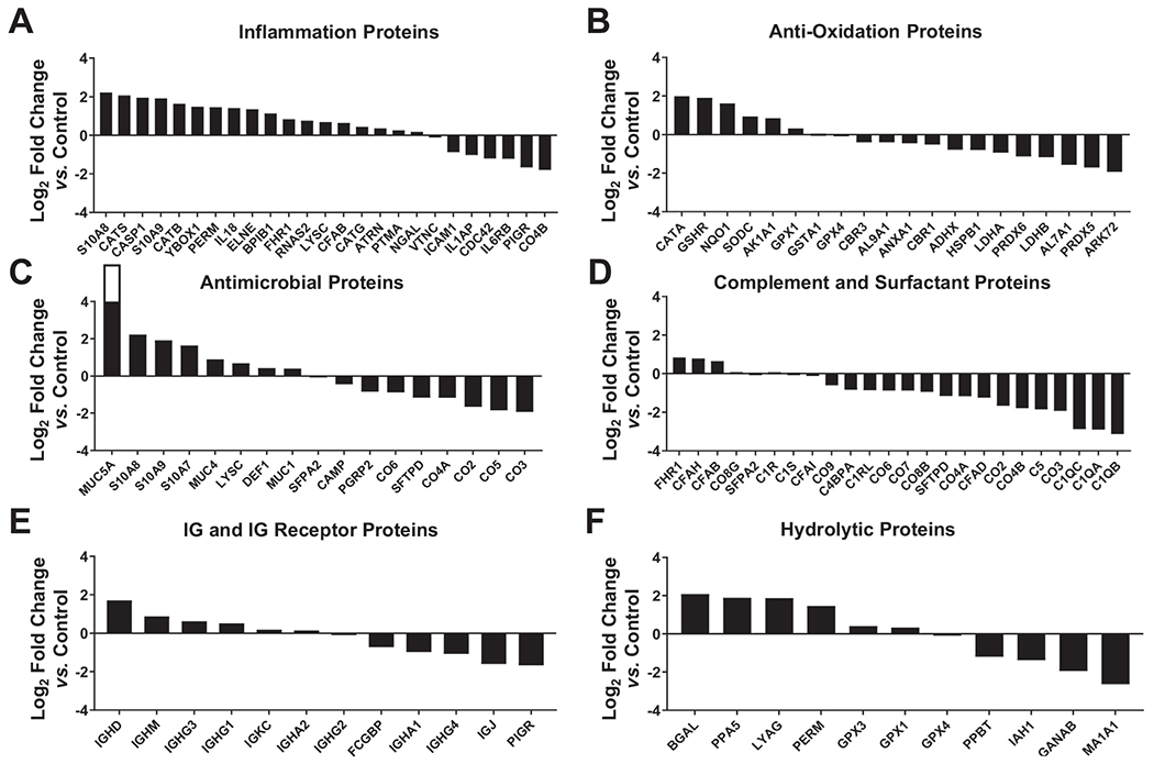
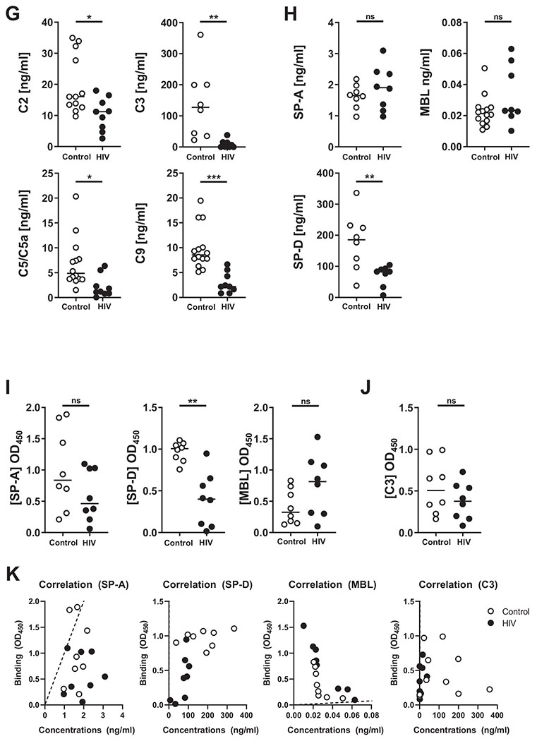
Fig. 2. Proteomic analyses and measurement of innate soluble mediators in ALF of both PLWH and control individuals.


Six major categories of ALF-derived proteins were identified: (A) inflammation proteins, (B) antioxidation proteins, (C) antimicrobial proteins, (D) complement and surfactant proteins, (E) IgG & IgG receptor proteins, and (F) hydrolytic enzymes or hydrolases. Differentially abundant proteins (DAPs) were identified by calculating their log2 fold-changes in the HIV-ALF relative to those in control-ALF. Each sample corresponds to ALF obtained from different human donors. For names of the proteins depicted and statistics, see supplemental material and Table S1 to S3. (G) Complement components: C2, C5/C5a, and C9; and (H) collectins SP-D and MBL were detected by Luminex. Complement component C3 and collectin SP-A were detected by ELISA. M.tb Erdman strain single-cell suspensions were exposed to control-ALF or HIV-ALF. Exposed-M.tb bacteria were washed, suspended in isotonic buffer, and plated onto 96-well plates. Monoclonal antibodies directed against (I) collectins SP-D, SP-A, MBL, and (J) complement component C3 were used to determine their amounts bound to M.tb. For assay controls, purified SP-D, SP-A, MBL, and C3 were used. Relative quantities of bound protein were quantified by standard ELISA by measuring the absorbance at OD450. (K) Correlations between the concentration levels and binding of SP-A, SP-D, MBL, and C3. Notice that regression line for SP-D and C3 overlaps with the y-axis. In G-K, each dot represents ALF from an individual subject. ALFs from control donors (n = 8-17) and PLWH subjects (HIV+, n = 7-10). Unpaired Student’s t test, *p < 0.05; **p < 0.005, ***p < 0.0005. Each sample corresponds to ALF obtained from different human donors.
