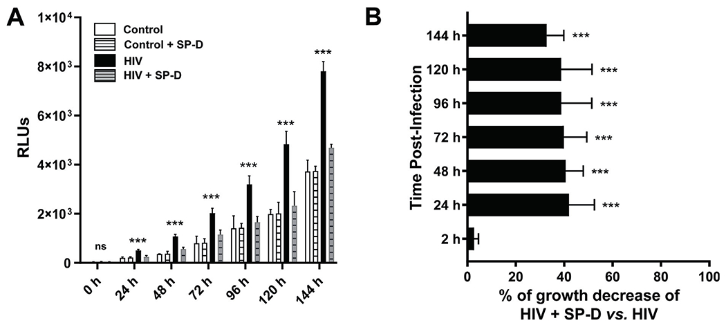
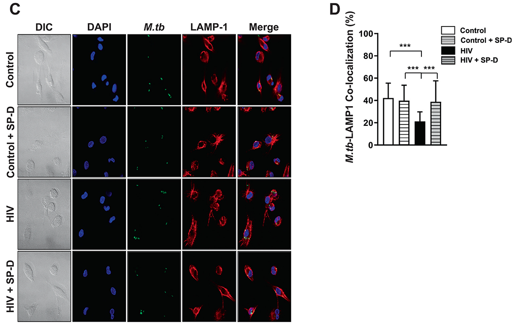
Fig. 4. Addition of SP-D to HIV-ALF restores macrophage control of M.tb growth.


(A) M.tb H37Rv-Lux was exposed to HIV-ALF, control-ALF, or to HIV-ALF or control-ALF each being supplemented with SP-D at its physiological concentration. Human macrophages were infected with these differently-exposed M.tb, and bacterial intracellular growth was measured in RLUs at different time points through 144 h. Representative experiment of n = 3, using ALF and macrophages in each case from different human donors. ANOVA Tukey-Posttest; HIV versus Control; ***p < 0.0005. (B) Overall data (n = 3) showing % of growth decrease for HIV-ALF + SP-D exposed M.tb versus HIV-ALF exposed M.tb. ***p < 0.0005. (C) GFP-M.tb Erdman was exposed to HIV-ALF, control-ALF, HIV-ALF+SP-D, or control-ALF+SP-D as described above prior to infection of macrophages on coverslips for 2 h (n = 3). P-L fusion events were visualized with confocal microscopy and enumerated by counting at least >150 independent events per coverslip. Shown are DIC, macrophage nucleus (DAPI, blue), phagosome containing GFP-M.tb (green), lysosomes (LAMP-1+, red), and P-L fusion events (co-localization, yellow). (D) Graph shows percentage of P-L fusion in terms of M.tb-LAMP-1 co-localization. Unpaired Student’s t test; Healthy versus HIV+; n = 3 ***p < 0.0005. Each sample corresponds to ALF obtained from different human donors.
