Fig. 3.
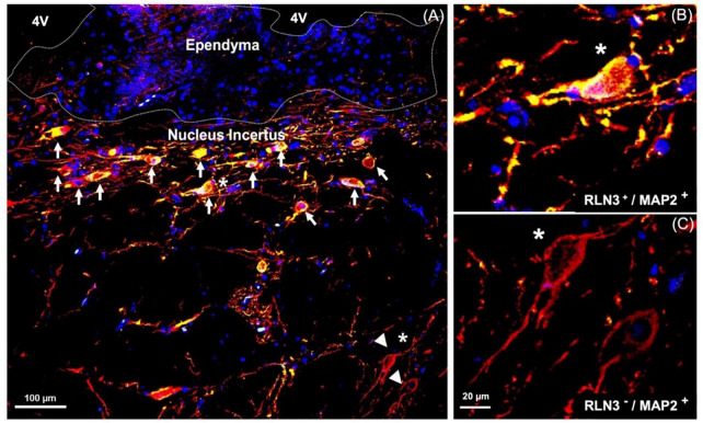
RLN3-immunoreactivity in neurons containing Microtubule-Associated Protein-2, a marker of neuronal cells, their perikarya, and dendrites [94]. A NI in human brain; white arrows indicate RLN3-immunoreactivity co-localized with MAP2. B Higher magnification view of a RLN3+/MAP2+ neuron indicated with a white star. C Higher magnification of RLN3−/MAP2+ neurons indicated in A with white arrowheads. MAP2 (red), RLN3 merging with MAP2 (yellow), DAPI (blue). Abbreviations: 4V, fourth ventricle, MAP2, microtubule-associated protein 2, RLN3, relaxin-3

