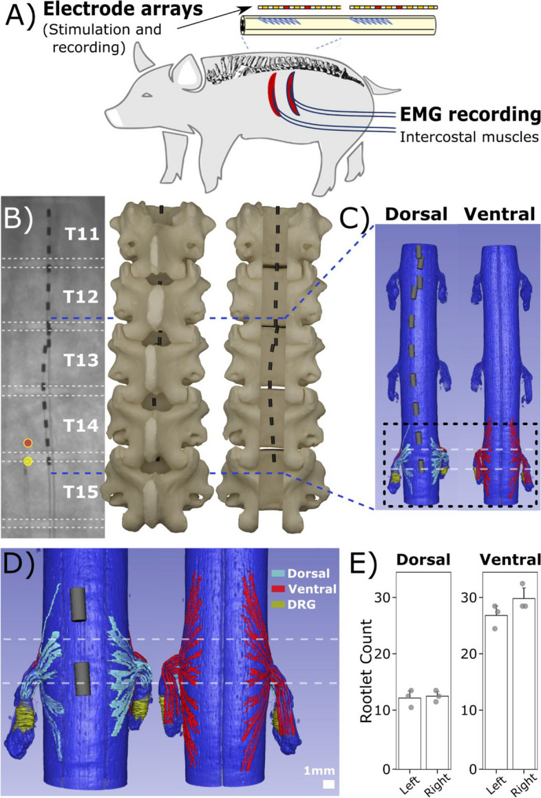
Fig. 1.

3D-model reconstruction of implanted epidural leads and spinal cord. A Experimental diagram. B For modeling purposes, x-ray images were taken from a representative subject’s (S0) thoracic vertebra segments with implanted epidural Octrode™ leads. Circular markers indicate the pair of electrode contacts used for SCS. Dashed white horizontal lines indicate intervertebral discs based on x-ray projections. 3D-models of the vertebra were generated to illustrate epidural lead placements with respect to the spinal column. C The subject’s spinal cord was reconstructed using microCT at levels around T12-T14 with epidural leads overlaid at ventral and dorsal viewing planes. D Reconstructed microCT segment of the spinal cord at T14 shows segmented dorsal and ventral rootlets and DRG. E Bilateral comparison of both dorsal and ventral rootlet counts (T12-T14) showed no significant differences in spinal rootlet count
