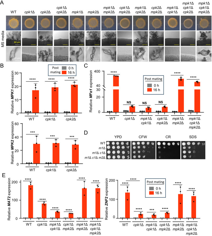
Fig 6.
Mpk1 and Mpk2 cooperate for the negative regulation of the Cpk1 MAPK pathway. (A) The MATa wild-type YL99a strain was cultured with the following MATα strains: wild type (H99), cpk1Δ, cpk2Δ, mpk1Δ, mpk2Δ, cpk1Δ cpk2Δ, mpk1Δ mpk2Δ, cpk1Δ mpk2Δ (YSB9206), mpk1Δ cpk2Δ (YSB8636), cpk2Δ mpk2Δ (YSB9584), mpk1Δ cpk1Δ (YSB6090), mpk1Δ cpk1Δ cpk2Δ (YSB10664), and mpk1Δ cpk1Δ mpk2Δ (YSB10643). Equal quantities of cells (107) were mixed and spotted onto the mating-inducing MS medium. After 11 days of incubation at room temperature in the dark, colony morphology, filamentation, and spore formation were observed under a brightfield microscope. (B) MATα wild-type, cpk1Δ, and cpk2Δ strains were subjected to unilateral mating with MATa wild-type on V8 medium. Following 0 and 16 h of incubation, RNA was extracted, cDNA was synthesized, and expression levels of MPK1 and MPK2 were quantified. (D) The wild-type and MAPK mutant strains were cultured and spotted under cell wall and membrane stress conditions using 0.05% CR, 0.5 mg/mL CFW, and 0.01% SDS. After 2 days, the plates were photographed. m1Δ refers mpk1Δ, m1Δ c1Δ refers mpk1Δ cpk1Δ, and m1Δ c1Δ m2Δ refers mpk1Δ cpk1Δ mpk2Δ. (C and E) MATα wild-type, cpk1Δ, mpk1Δ cpk1Δ, and mpk1Δ cpk1Δ mpk2Δ strains underwent unilateral mating with the MATa wild-type YL99a strain. Post 0 and 16 h of incubation, RNA was extracted, and cDNA was synthesized. Expression of MFα1, MAT2, and ZNF2 was quantified by quantitative reverse transcription-PCR (qRT-PCR) with three biological replicates. Statistical significance of the data was determined using one-way ANOVA with Tukey’s multiple-comparison test: *, P < 0.05; **, P < 0.01; ***, P < 0.001; ****, P < 0.0001; NS = not significant.

