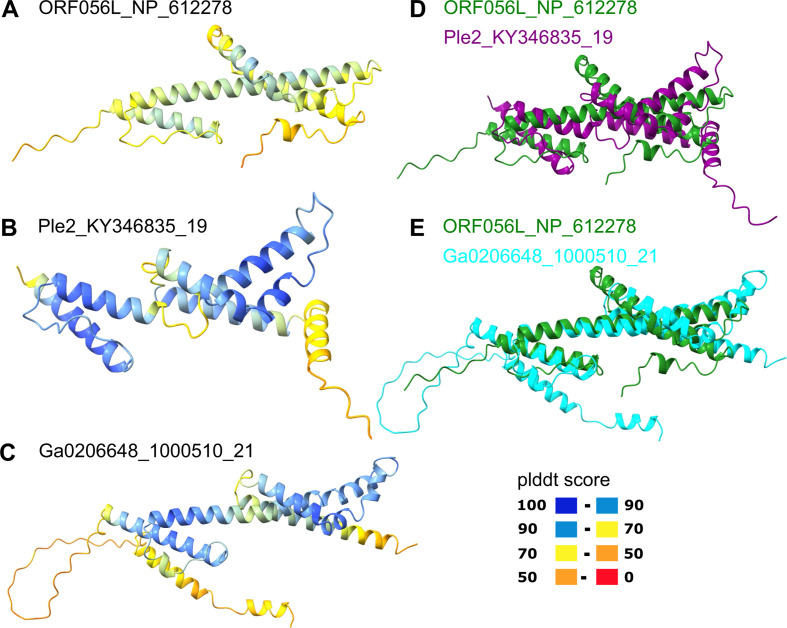
Fig 7.
Comparison of the structural models of Mriya_48 protein and iridovirus envelope protein. (A) Iridovirus enveloped protein (ORF056L; NP_612278); (B) Ple2_KY346835_19; (C) Ga0206648_1000510_21; (D) Superposition of ORF056L (green) and Ple2_KY346835_19 (purple); (E) Superposition of ORF056L (green) and Ga0206648_1000510_21 (cyan). In panels A–C, the structures are colored according to the plddt score.

