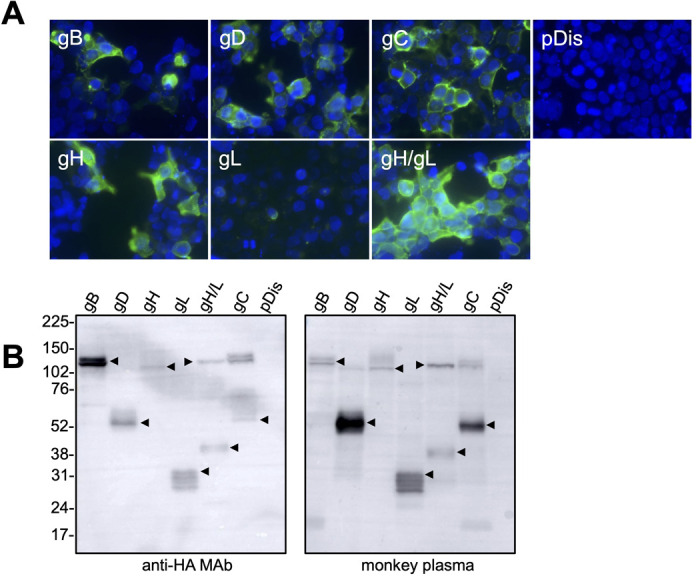
Fig 1.

Expression of B virus glycoproteins. (A) 293T cells were transfected with plasmids encoding BV glycoproteins. After a 48– to 72-h incubation, the cells were fixed with 10% formalin and were stained with an anti-HA monoclonal antibody detected with a secondary Alexa-Fluor-488-conjugated antibody. (B) The plasmid-transfected cells were also analyzed by Western blotting using an anti-HA monoclonal antibody and anti-BV-antibody-positive macaque plasma. Molecular weight marker labels (kDa) are shown on the left, and the protein of interest in each lane is indicated by an arrow.
