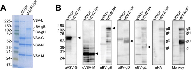
Fig 3.

Incorporation of BV glycoproteins into VSV pseudotype particles. Pseudotyped VSV∆G/Luc bearing the BV glycoproteins gB, gD, gH, and gL (VSV/BVpv), and VSV∆G/Luc-*G (VSVpv) were partially purified by ultracentrifugation in 25% sucrose medium, separated by SDS-PAGE, and analyzed by Coomassie blue staining (A) or by Western blotting (B) using the indicated antibodies. Monkey, BV-positive macaque plasma. Molecular weight marker labels (kDa) are shown on the left, and the proteins of interest in each lane are indicated by an arrow or a label.
