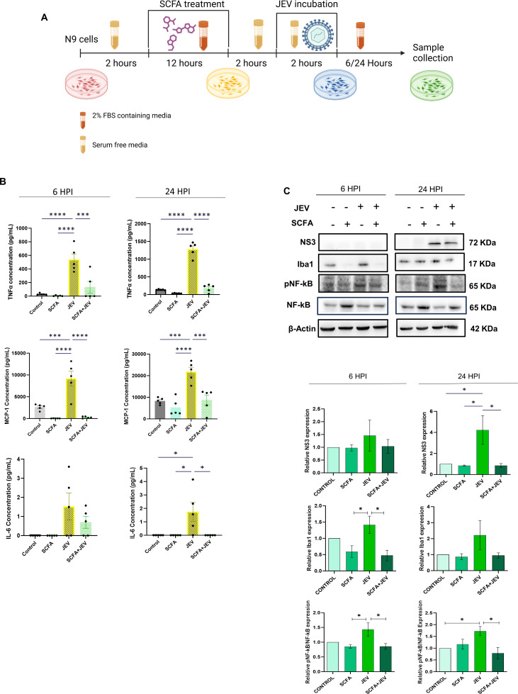
Fig 1.
SCFA pretreatment reduces expression of inflammatory markers post-JEV infection in microglial cells. (A) Schematic representation of the experimental paradigm followed. (B) Cells supernatant isolated form N9 microglial cells post SCFA/PBS pretreatment and/or JEV infection (multiplicity of infection [MOI] 3 ) were subjected to cytokine bead analysis. Graphs on the upper panel (blue dot) represent cytokine expression 6 HPI and those on the lower panel (orange dot) represent 24 HPI. Data presented as absolute concentration (µg/mL) ± SEM. (C) Representable immunoblots from cell lysate showing various inflammatory markers at 6 HPI and 24 HPI post SCFA pretreatment and JEV infection. (D) Bar plots showing densitometric quantification of the immunoblots in panel B. Data represented as mean fold change ± SEM with respect to control from a minimum of three independent experiments. P-values were determined (*, P < 0.05; **, P < 0.01; ***, P < 0.001) using one-way analysis of variance and Tukey’s post hoc correction.

