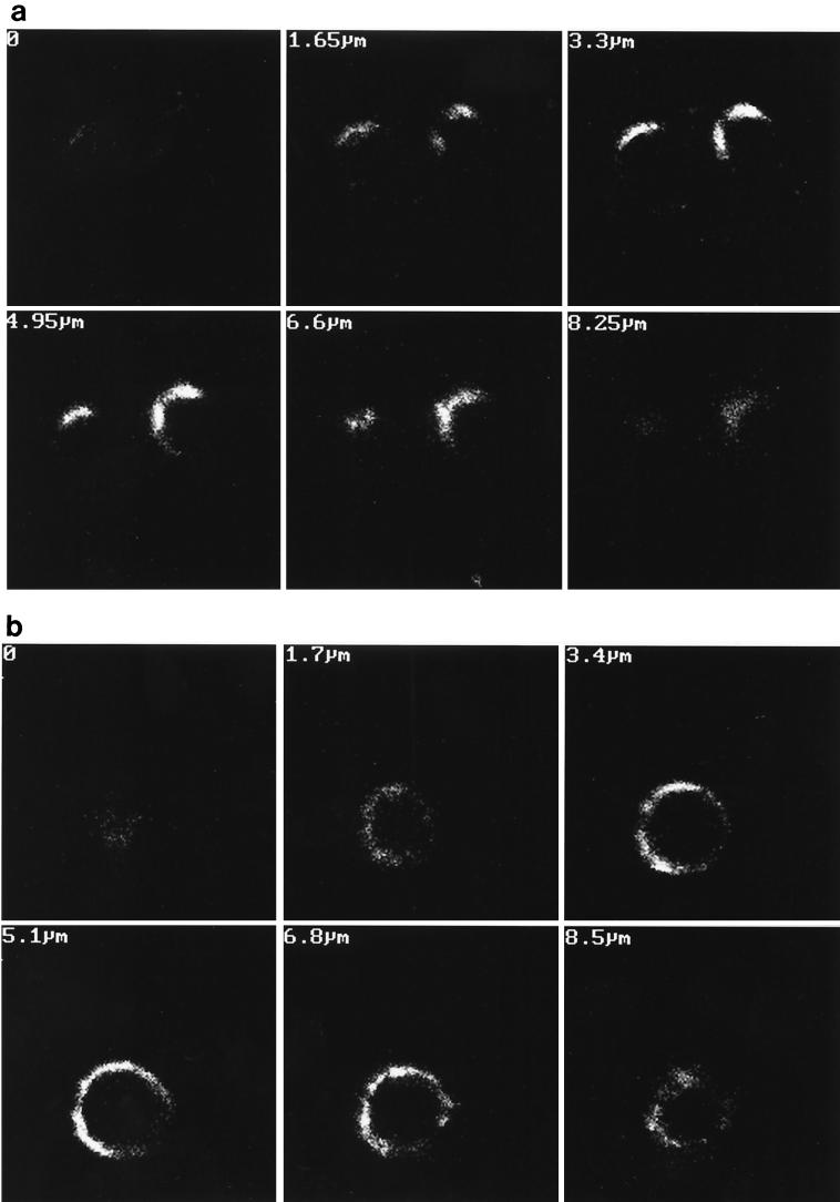
FIG. 3.
Polarized localization of p24. Transfected Jurkat cells at the peak of viral replication (9 days posttransfection) were analyzed by confocal microscopy with a monoclonal anti-p24 antibody as described in Materials and Methods. Cells transfected with wild-type proviral DNA exhibit polarized localization of the p24 protein (a), while cells transfected with mutant Y712S DNA showed a lack of polarization (b). Numbers in top left corners indicate the depth of each serial section. Magnification, ×360.

