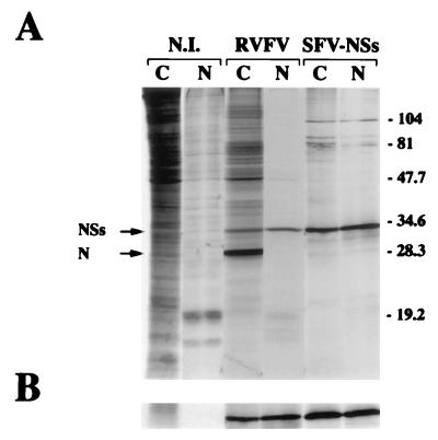
FIG. 2.
Distribution of the NSs protein between the cytoplasmic and nuclear fractions. (A) Confluent monolayers of BSR cells were infected with the MP12 strain of RVF virus or SFV-NSs or mock infected (N.I.). At 6 h postinfection, the cells were labeled with 200 μCi of a mixture of [35S]methionine and [35S]cysteine per ml for 1 h and fractionated into cytoplasmic (C) and nuclear (N) extracts. The proteins were analyzed by electrophoresis in SDS–12% polyacrylamide gels and autoradiography. The positions of the N and NSs proteins and the molecular mass markers (in kilodaltons) are shown on the left and right, respectively. (B) Nuclear and cytoplasmic extracts were immunoprecipitated with the mouse polyclonal antibodies against the NSs protein. The immune complexes were analyzed by SDS–12% polyacrylamide gel electrophoresis followed by autoradiography.

