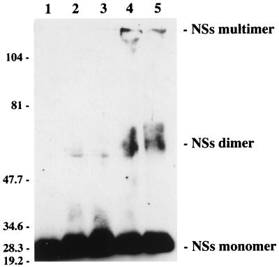
FIG. 6.
Analysis of nucleoplasmic proteins after cross-linking with various concentrations of glutaraldehyde. Proteins from the nucleoplasmic fraction were treated with 0% (lane 1), 0.05% (lane 2), 0.1% (lane 3), 0.5% (lane 4), or 1% (lane 5) glutaraldehyde, electrophoresed in an 8% polyacrylamide gel, and analyzed by Western blotting with NSs-specific antibodies. The positions of the molecular size markers in kilodaltons are shown on the left.

