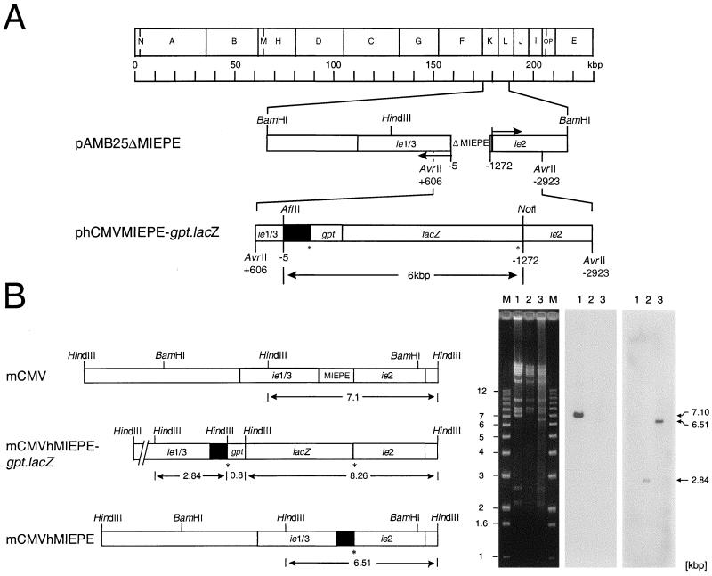
FIG. 1.
Construction and verification of recombinant viruses. All map positions are given relative to the 5′ start site (counted as +1) of the ie1/3 transcription unit of mCMV, and illustrations are drawn to scale. (A) Map of plasmid constructs for homologous recombination. The HindIII physical map of the mCMV Smith strain genome is shown at the top. For the construction of the mCMV MIEPE deletion plasmid pAMB25ΔMIEPE, plasmid pAMB25 was digested with MluI and MunI and surplus deletions were restored by PCR, resulting in a final deletion of 1,267 bp (ΔMIEPE). Arrows indicate the orientations of ie1/3 and ie2 transcription. Plasmid phCMVMIEPE-gpt.lacZ was generated by insertion of the hCMV enhancer and core promoter (solid box, representing hCMV nucleotides −14 to −601 relative to the start site of the hCMV ie1-ie2 transcription unit) and of a gpt.lacZ reporter gene cassette flanked by loxP sites (asterisks). (B) MIEPE swap mutants. Maps are shown on the left and the corresponding HindIII cleavage analysis is shown on the right. (Left) Expanded HindIII fragments K and L of the mCMV Smith strain genome are shown on the top, illustrating the location of the MIEPE and flanking ie sequences within the authentic 7.1-kbp L fragment (map corresponding to lane 1). Replacement of the mCMV MIEPE by the hCMV MIEPE (solid box) and insertion of a gpt.lacZ reporter gene cassette flanked by loxP sites (asterisks) created novel HindIII fragments of 0.8, 2.84, and 8.26 kbp in the genome of recombinant virus mCMVhMIEPE-gpt.lacZ (map corresponding to lane 2). For the generation of recombinant virus mCMVhMIEPE, the gpt-lacZ cassette was removed from mCMVhMIEPE-gpt.lacZ via Cre recombinase-mediated loxP-specific recombination, leaving a single loxP site (asterisk) and generating a shortened HindIII L fragment of 6.51 kbp (map corresponding to lane 3). (Right) Purified virion DNA was subjected to cleavage by HindIII, and fragments were analyzed by agarose gel electrophoresis, Southern blot, and hybridization with MIEPE type-specific γ-32P-end-labeled oligonucleotide probes. Lanes: M, indicated size markers; 1, DNA of parental virus mCMVΔorf152; 2, DNA of mCMVhMIEPE-gpt.lacZ; 3, DNA of mCMVhMIEPE. Left panel, ethidium bromide-stained gel; center panel, autoradiograph obtained after hybridization of the Southern blot with the 30-bp probe mE-oligo-P; right panel, autoradiograph obtained after stripping of the same filter followed by hybridization with the 30-bp oligonucleotide probe hE-oligo-P. See Fig. 3 for the map locations of the two probes.

