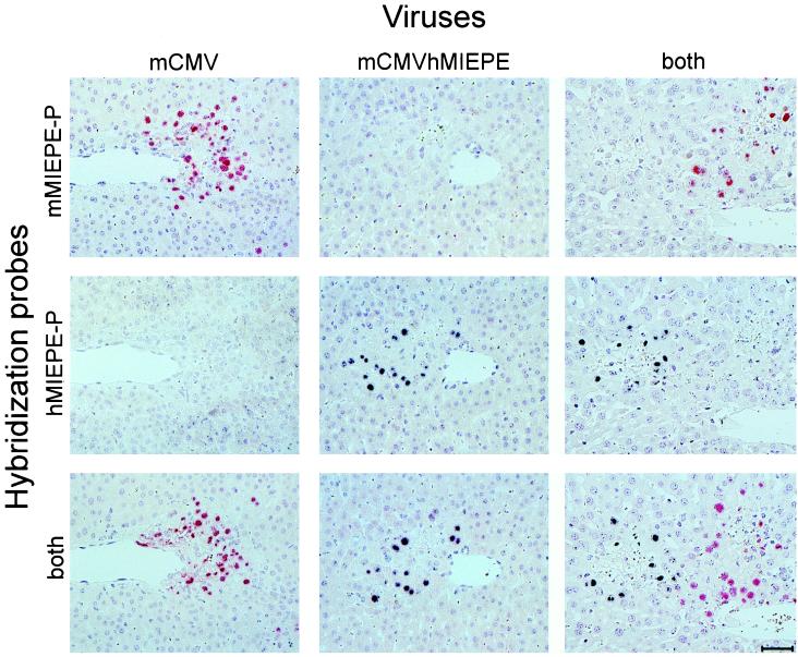
FIG. 4.
Two-color MIEPE type-specific ISH in liver tissue sections. After immunoablative treatment, groups of BALB/c mice were infected intravenously with either mCMV or recombinant virus mCMVhMIEPE or were coinfected with both viruses. Histological analysis was performed on day 9 after infection. Three serial sections, sharing a central vein as a landmark, were selected for hybridization. The first section of each series was hybridized with a polynucleotide probe specific for the MIEPE of mCMV (mMIEPE-P; red staining), the second was hybridized with a polynucleotide probe specific for the MIEPE of hCMV (hMIEPE-P; black staining), and the third was hybridized with a mixture of both probes. See Fig. 3 for the map locations of the two probes. Results are documented for all nine possible combinations. Counterstaining was performed with hematoxylin. The bar in the bottom right panel represents 50 μm.

