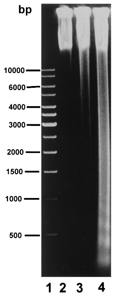
FIG. 2.
DNA fragment analysis of CHSE-214-EGFP cells infected with IPNV E1-S (MOI of 1). DNA was isolated (as described in Materials and Methods) from uninfected CHSE-214 cells as a negative control after 0 (lane 2) and 8 (lane 3) h of incubation and from cells infected for 8 h with an MOI of 1 of E1-S (lane 4), electrophoresed through 1.2% agarose gels, and visualized by ethidium bromide staining. Lane 1 contained molecular size markers (1-kb DNA ladder from USA MBI Fermentas Inc. for sizing of linear fragments ranging from 500 bp to 1 kb).

