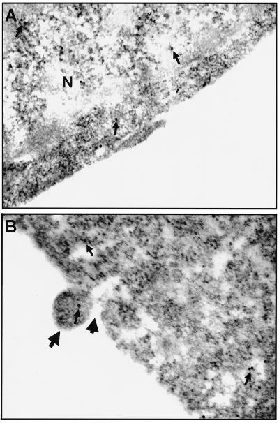
FIG. 5.
Immunoelectron micrographs of ultrathin sections of CHSE-214-EGFP cells that were uninfected or infected with IPNV and labeled with anti-GFP IgG. (A) Normal CHSE-214-EGFP cell used as a negative (N) control on which labeled EGFP is present (arrows) and EGFP formed dimers. (B) CHSE-214-EGFP cell infected with IPNV (MOI of 1) at 8 h p.i. upon which labeled EGFP is present (small arrows). Nontypical apoptotic morphological changes were observed at this pre-late apoptotic cell stage such as the formation of MV (large, long arrow) and, finally, the MV pinching off from the plasma membrane of the apoptotic cell (large, short arrow).

