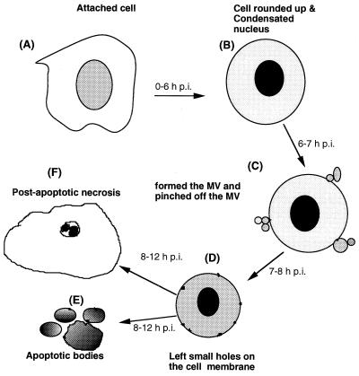
FIG. 7.
Diagram illustrating the morphological changes induced in fish cells by IPNV infection. (A) Normal attached cell. In the early stage of apoptosis, the cell detaches from the extracellular martix (A to B, 0 to 3 h p.i.). In the middle stage, the apoptotic cell is rounded up (A to B, 3 to 6 h p.i.). To enter this pre-late apoptotic stage (B to C, 6 to 7 h p.i.), there is a rapid process which follows that includes MV formation and MV pinching off from the plasma membrane. In the middle stage, the apoptotic cell is left with small holes in the cell membrane (C to D, 7 to 8 h p.i.). Finally, in the late apoptotic stage, either membrane-bound apoptotic bodies (D to E, 8 to 12 h p.i.) are formed or a postapoptotic necrosis process occurs (D to F, 8 to 12 h p.i.) in which the condensed chromatin encloses the nuclear membrane.

