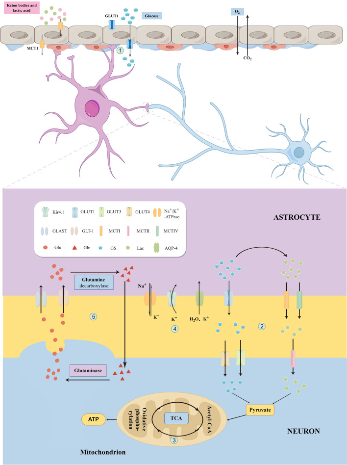
Fig. 2.
Synaptic physiologic activity of neurons and astrocytes in cortical spreading depression (CSD). Glucose transporter 1 (GLT1) is primarily expressed in the endothelial cells and astrocytes. Glucose enters astrocytes via GLT1 through the blood–brain barrier (Step 1) and undergoes oxidative metabolism to produce lactate (Step 2). Lactate enters the neurons through a monocarboxylate transporter (MCT) to produce pyruvate, which undergoes oxidative decarboxylation to form acetyl coenzyme A, entering the tricarboxylic acid cycle (Step 3). Synaptic gap excess K+ influxes into astrocytes during CSD (Step 4), leading to swelling of astrocyte endfeet. The massive release of glutamate (Glu) from neurons during CSD enters astrocytes through Glu transporter 1 (GLT-1) and Glu aspartate transporter (GLAST) to generate glutamine (Gln). Gln leaves astrocytes by exocytosis, enters the neuron by endocytosis, and is converted to Glu for its release into the synaptic gap in a process known as the Glu–Gln cycle (Step 5)

