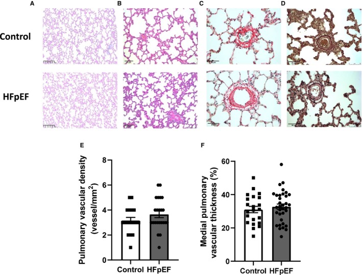
Figure 2. Pulmonary artery morphometry in HFpEF rats.

Representative slides of hematoxylin–eosin‐stained pulmonary (A; scale bar: 250 μm) and pulmonary artery (B; scale bar: 75 μm) sections, Picrosirius Red‐ (C; scale bar: 50 μm) and orcein‐ (D; scale bar: 50 μm) stained pulmonary artery sections. PicroSirius Red staining was performed to detect fibrotic areas, with collagen fibers stained in red and orcein staining to detect internal and external elastic lamellae. Vascular density (E; expressed as the number of pulmonary arteries/mm2 lung parenchyma) and morphometry on pulmonary arterioles (with external diameter <150 μm) expressed as medial wall thickness percentage (%MT) vs external diameter (F) in lungs of control (white bars; n=9) and HFpEF (black bars; n=13) rats. Values are expressed as mean±SEM. HFpEF indicates heart failure with preserved ejection fraction.
