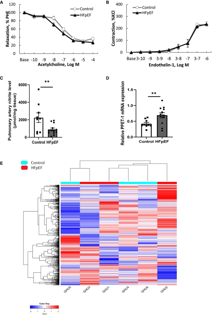
Figure 3. Ex vivo pulmonary artery vasoactive assessment and pulmonary pathobiological evaluation, including RNA sequencing in HFpEF rats.

Concentration‐response curves to acetylcholine (A; tested from 10−10 to 10−4 mol/L−1) after phenylephrine (PHE) precontraction and to endothelin‐1 (B; tested from 10−8 to 10−3 mol/L−1) in endothelium‐intact pulmonary artery segments from control (white circles; n=9) and HFpEF (black triangles; n=13) rats. Concentration of nitrites in supernatants of endothelium‐intact pulmonary artery rings from control (white bars; n=9) and HFpEF (black bars; n=13) rats and incubated during 1 hour in Krebs solution (C). Relative mRNA expression of pre‐proendothelin‐1 (PPET‐1) in lungs from control (white circles; n=9) and HFpEF (black triangles; n=13) rats (D), Values are expressed as mean±SEM. ** 0.001<P<0.01, HFpEF vs control rats. Heatmap (E) showing nonclustering of genes from RNA sequencing in 3 randomly chosen control (in blue) and in three randomly chosen HFpEF (in red) rats. Raw Z scores are shown on the heatmap. HFpEF indicates heart failure with preserved ejection fraction.
