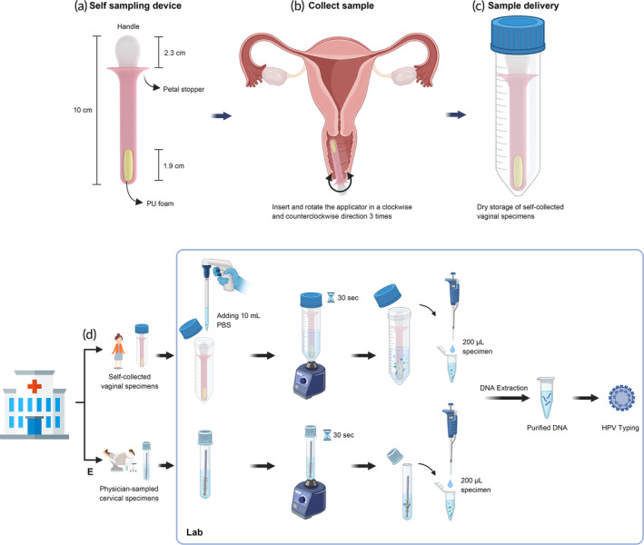
FIGURE 1.

Schematic diagrams of various components and procedures related to the self‐sampling device and sample processing procedures in this study. (a) The self‐sampling device provided to participants for the study is approximately 10 cm long, with a comfortable grip handle measuring 2.3 cm and a polyurethane foam sample collection pad embedded in the distal end. (b) Participants were provided instructions on how to perform the self‐collection procedure during their visit to the outpatient clinic. (c) Specimens were stored in a sterilized 50 mL centrifuge tube in a dry state for transportation to the laboratory for further processing and analysis. (d) Initially, a self‐collected vaginal specimen was mixed with 10 mL of PBS buffer and then vortexed for 30 s to wash cells from the foam. From this, 200 μL of the specimen was transferred into a microtube and subjected to DNA extraction protocol. (e) In the case of physician‐sampled cervical specimens, the cells from the brush were washed by vortexing for 30 s. From this, 200 μL of the specimen was then transferred into a microtube and subjected to the DNA extraction protocol. Both purified DNA samples were subjected to HPV genotyping using the DR. HPV Genotyping IVD Kit, following the manufacturer's instructions. The diagram was created using BioRender.com (https://app.biorender.com/).
