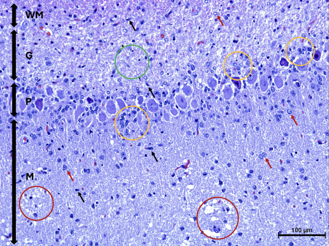
FIGURE 2.

Microscopic image of the cerebellar cortex of Case 1 stained with Hematoxylin and Eosin. The granular cell layer (G) is displaying marked pallor because of hypocellularity of the granule cell neurons (green circle). There are Bergmann glial cells (yellow circles) located adjacent to the Purkinje cell layer (P). The molecular layer (M), is displaying mild astrogliosis (red arrows) including the presence of microglia (black arrows) and spongiosis (red circles). White matter (WM).
