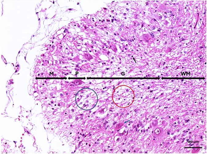
FIGURE 4.

Microscopic image of the cerebellar cortex of Case 2 stained with Hematoxylin and Eosin. The granular cell layer (G) is displaying marked hypocellularity of the granule cell neurons with marked vacuolation of the neuropil (red circle), as well as shrunken pyknotic granular cell neurons (black arrow). There is rare loss of Purkinje neurons (blue circle) as well as rare shrunken angular hypereosinophilic Purkinje neurons (blue arrow) in the Purkinje cell layer (P). The molecular layer (M) size is reduced. White matter (WM).
