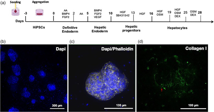
FIGURE 2.

HiPSCs differentiation protocol in Biomimesys® Liver for the generation of liver organoids. (a) Schematic diagram of the hiPSC differentiation protocol into liver organoids. (b) DAPI staining of liver organoids at low magnification (overall view of a 96‐well plate). (c) Actin filament staining in liver organoids using phalloidin‐488 (green). (d) Collagen type I fibers (blue) visualized using bi‐photonic microscopy in a liver organoid.
