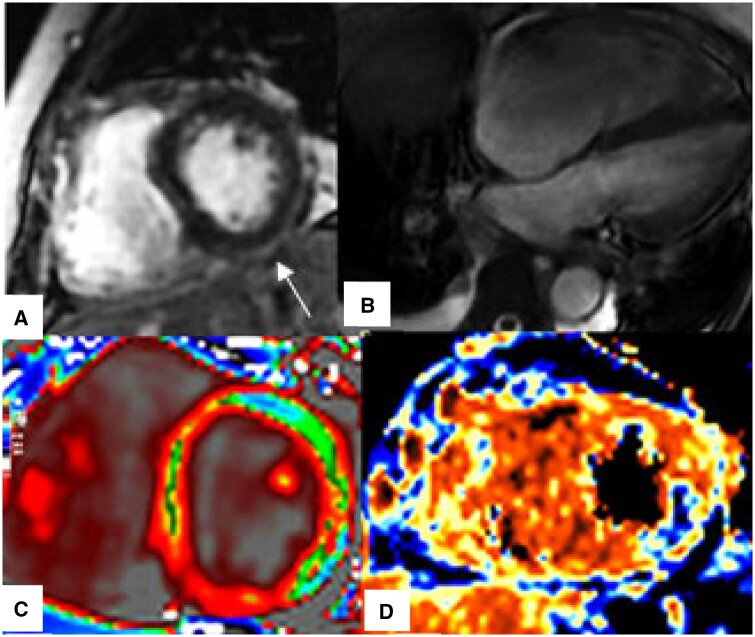
Figure 4.
CMR findings of myocardial injury in patients with acute severe COVID-19. (A) LGE, Non-ischaemic subepicardial enhancement involving inferior and inferolateral segments (arrow), (B) Four-chamber SSFP, Dilated right ventricle, (C) T1 map, Diffusely increased T1-relaxation times, (D) T2 map, Focal high T2-values in the mid inferolateral wall (blue colour).

