
Figure 3.
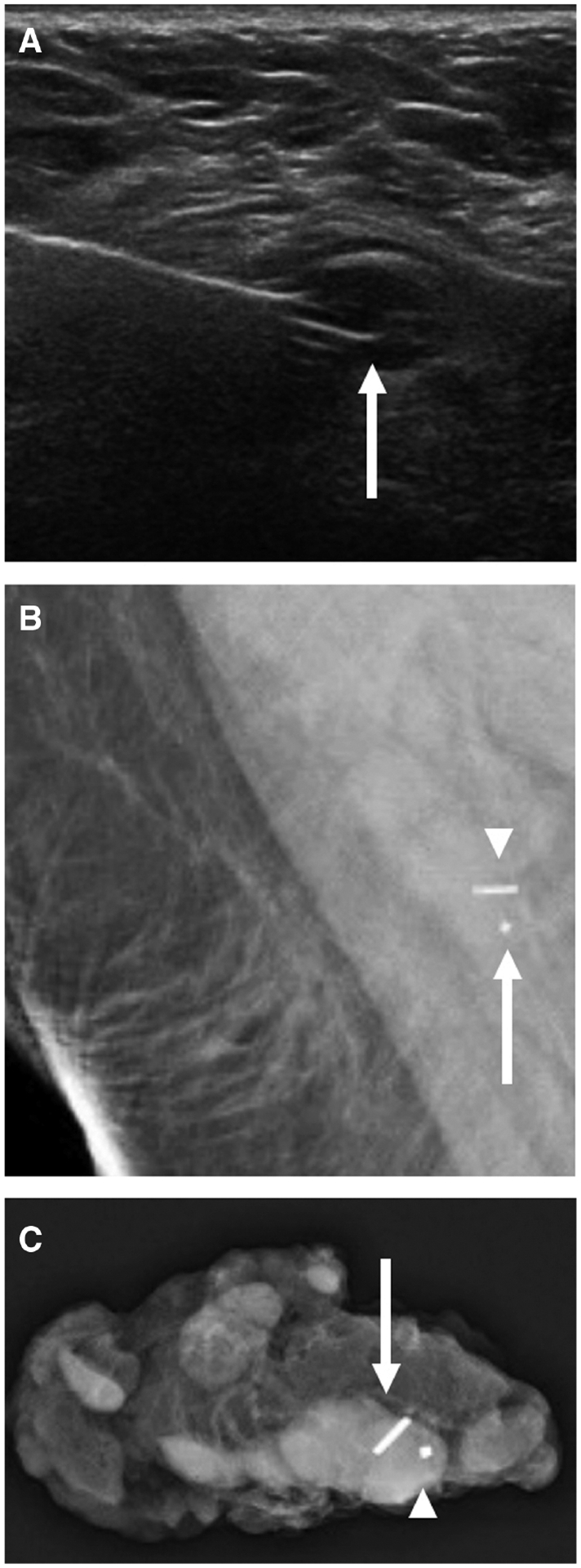
Images of a 48-year-old woman with biopsy-proven right breast invasive ductal carcinoma with ductal carcinoma in situ, metastatic to the right axilla, status post-neoadjuvant chemotherapy (same patient in Figure 2). A: US-guided radioactive seed localization of a biopsy-proven metastatic level 1 right axillary lymph node, with the tip of the needle deploying the seed in the lymph node (arrow). Post-procedural right breast mammogram coned down to the axilla (B) shows the seed (arrowhead) immediately adjacent to the biopsy clip (arrow). Specimen radiograph of the right axilla (C) shows the metastatic lymph node containing the marker (arrowhead) and seed (arrow).