
Figure 4.
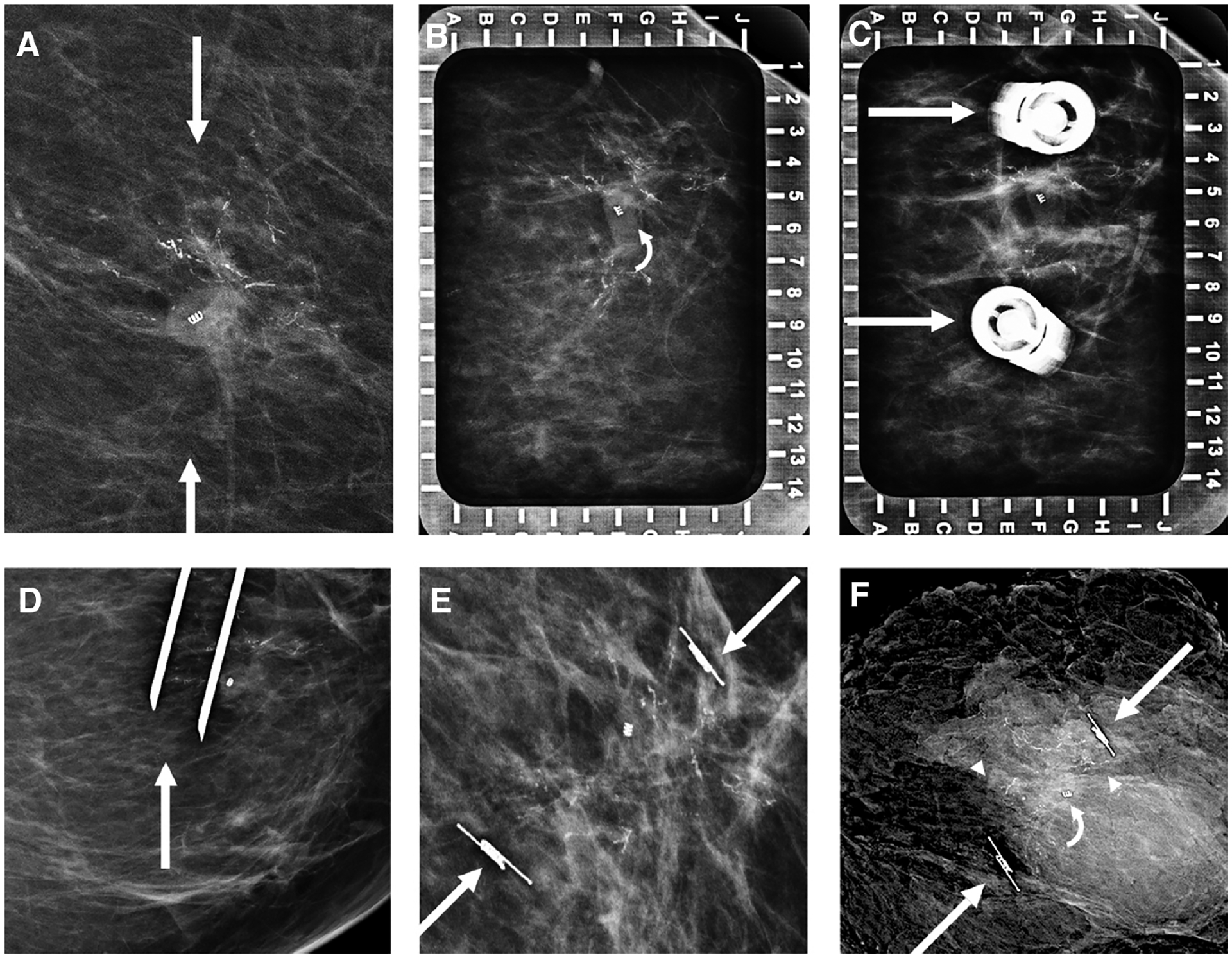
Images of a 43-year-old woman with biopsy-proven left breast ductal carcinoma in situ (DCIS). A: Bracketing SAVI SCOUT localization targeting residual calcifications (arrows) surrounding an open coil biopsy marker was requested. Alphanumeric grid (B) for bracketing from a superior approach shows residual calcifications surrounding the open coil biopsy clip (curved arrow). Repeat imaging (C) shows the SAVI SCOUT needle hubs (arrows) at the medial and lateral edges of residual calcifications. Orthogonal mediolateral projection (D) confirms the needle tips (arrow) to be approximately 6 mm distal to the inferior residual calcifications just prior to SAVI SCOUT deployment. Craniocaudal mammogram (E) confirms accurate placement of the SAVI SCOUT devices (arrows) bracketing the residual calcifications and biopsy marker. Specimen radiograph (F) shows residual calcifications (arrowheads), the biopsy marker (curved arrow), and SAVI SCOUT devices (arrows). At final pathology, the patient was upgraded to invasive ductal carcinoma, not otherwise specified, with residual DCIS.