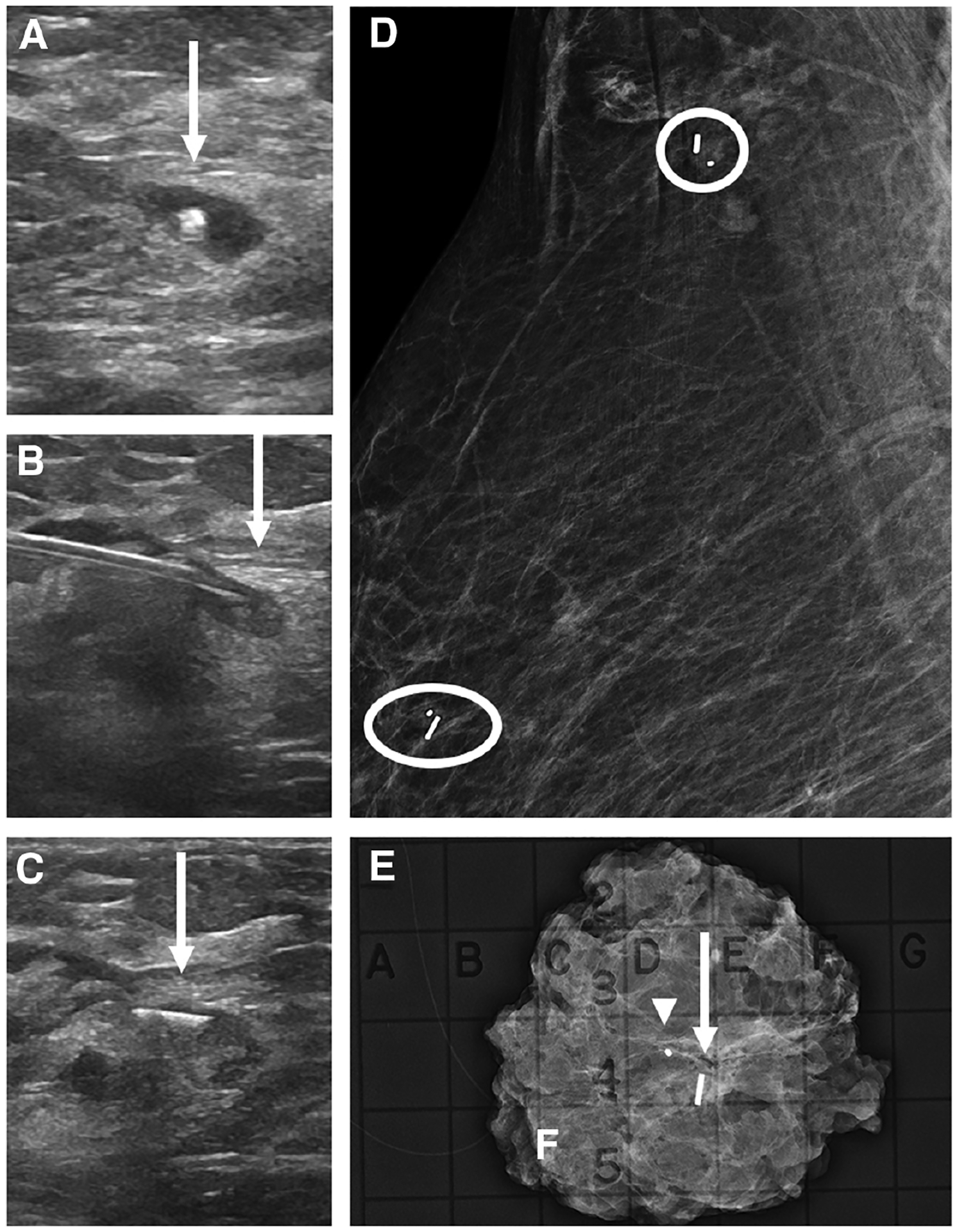
Figure 5.

Images of a 66-year-old woman with biopsy-proven right breast invasive ductal carcinoma with lobular features, metastatic to the right axilla, presenting for breast and axillary Magseed localization following neoadjuvant chemotherapy. Right breast US (A) shows a residual mass in the right breast with a central biopsy clip (arrow). Right breast US (B) shows the Magseed needle with the tip in the residual mass (arrow) prior to deployment. Following deployment, US (C) shows the Magseed within the superior edge of the residual mass (arrow). Post-procedural right breast mediolateral oblique mammogram (D) shows the Magseed adjacent to the biopsy clip in the upper breast (oval). A second Magseed was also placed under US guidance at the biopsy-proven level 1 axillary lymph node containing a biopsy marker (circle). Specimen radiograph of the right breast (E) shows the marker (arrowhead), Magseed (arrow), and residual surrounding focal asymmetry. The axillary specimen was not submitted for review.
