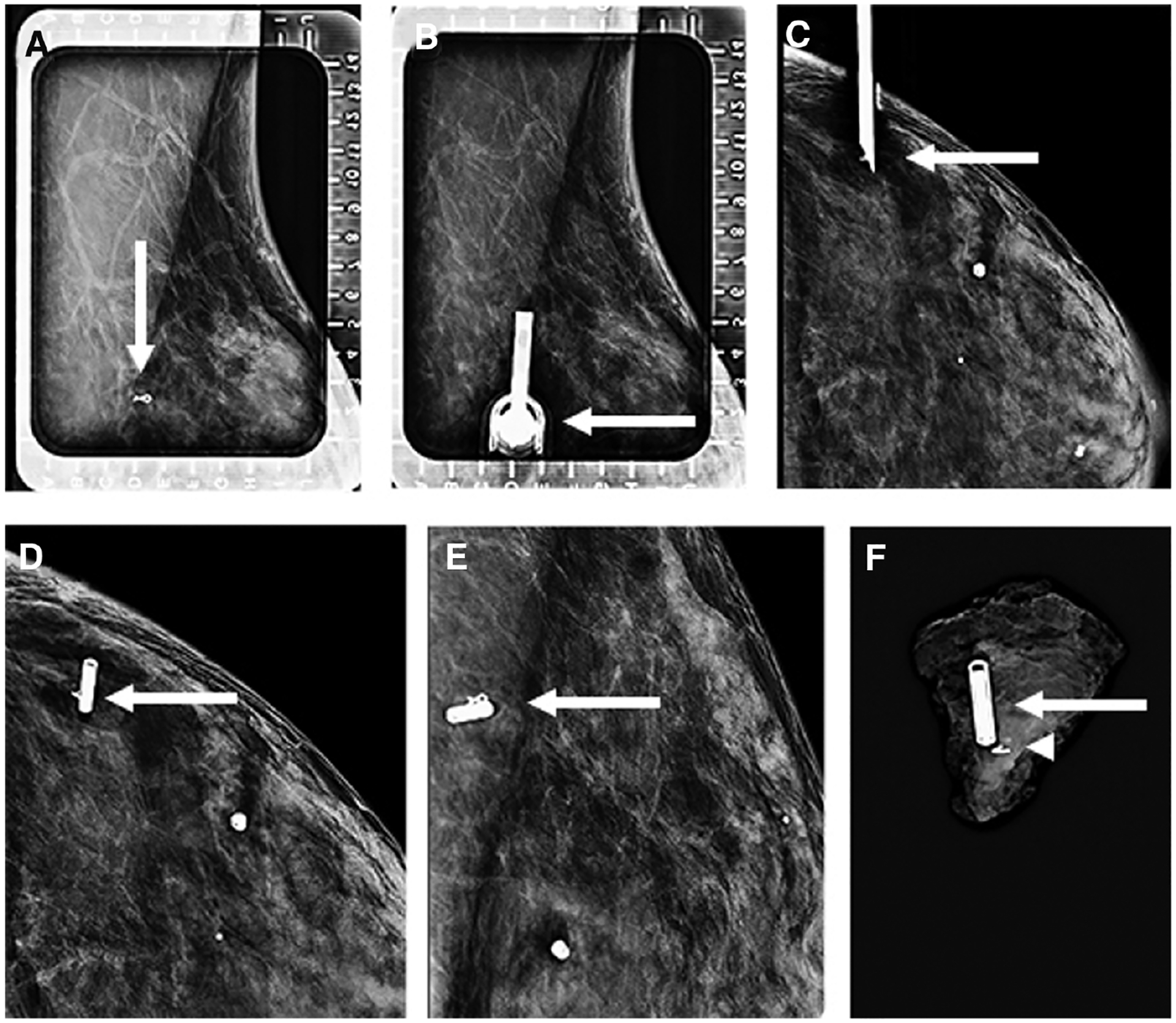
Figure 6.

Images of a 44-year-old women status post-neoadjuvant therapy for left breast invasive ductal carcinoma, not otherwise specified, who presented for radiofrequency identification (RFID) tag localization of a ribbon clip marking the site of the treated cancer. Left breast alphanumeric grid for localization from a lateral approach (A) demonstrates the ribbon clip (arrow). Repeat image (B) shows the needle housing the RFID tag inserted with the hub (arrow) directly overlying the ribbon clip. Orthogonal craniocaudal (CC) view (C) demonstrates the needle tip (arrow) at the depth of the ribbon clip before deployment. The RFID tag was subsequently deployed, with the CC (D) and mediolateral (E) views demonstrating the RFID tag (arrow) to be adjacent to the ribbon clip. Specimen radiograph (F) confirms that the ribbon clip (arrowhead) and RFID tag (arrow) are within the surgical specimen.
