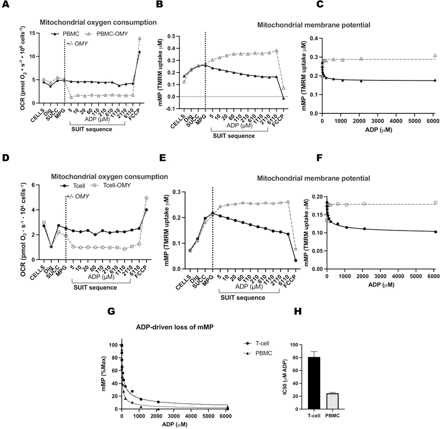
Figure 4: ATP-synthase drives ADP-driven decrease in membrane potential in T-cells and PBMCs.

(A-H) The protocol described here was tested in PBMCs and T-cells. Two O2K chambers were injected with PBMCs, and two chambers of an additional O2K were injected with T-cells from the same participant. After injecting substrates malate, pyruvate, and glutamate in all chambers, one chamber of PBMCs and T-cells received oligomycin. Oligomycin prevented any ADP-driven rise in respiration in (A) PBMCs and (D) T-cells or decline in mitochondrial membrane potential in (B,C) PBMCs and (E,F) T-cells. (G,H) ADP sensitivity was greater in PBMCs compared to T-cells.
