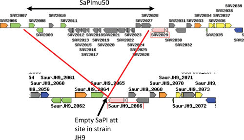
FIGURE 2.

SaPI genomic pattern. At the top is the groEL region of S. aureus strain mu50, with a classical SaPI inserted at the 3′ end of groEL. The basic SaPI genes are in gray, and accessory genes at the left end are in green. Note the typical transcriptional divergence. Below is shown the corresponding region of strain JH9, in which the SaPI att site at groEL is empty.
