Figure 3.
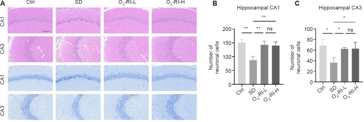
O3-RI improves the hippocampal pathology in SD mice.
Note: (A) The images of hematoxylin and eosin (upper two rows) and Nissl (lower two rows) staining of hippocampal CA1 and CA3 regions. Compared with the Ctrl group, the neurons in the SD group were necrotic and damaged, which was mainly manifested by the shrinkage and condensation of the nucleus, the enhancement of basophilia, and the number of neurons was also significantly reduced, which was gradually improved in the two O3-RI treatment groups. Scale bars: 100 μm. (B) Quantity of neurons in the hippocampal CA1 (B) and CA3 (C) regions using Nissl staining. Data are expressed as mean ± SD (n = 6). *P < 0.05, **P < 0.01 (one-way analysis of variance, followed by Tukey's multiple comparison test). Ctrl: Control; ns: no significance; O3-RI: ozone rectal insufflation; O3-RI-H: high-concentration O3-RI; O3-RI-L: low-concentration O3-RI; SD: sleep deprivation.

