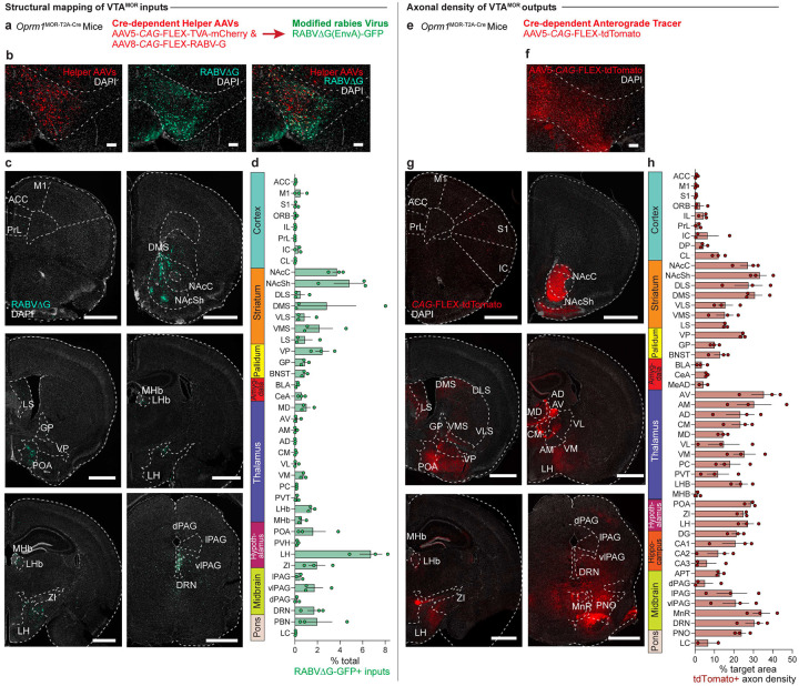
Figure 3: Structural input-output mapping of VTAMOR neurons.
(a) Experimental schematic for rabies-mediated monosynaptic labelling of VTAMOR inputs in Oprm1MOR-T2A-Cre mice through a unilateral injection of AAVs expressing Cre-dependent TVA-mCherry (AAV5-CAG-FLEX-TVA-mCherry) and rabies glycoprotein (AAV8-CAG-FLEX-RABV-G) helper viruses in the VTA, followed by a subsequent injection of the EnvA-pseudotyped, G-deleted, GFP-expressing rabies virus (RABVΔG-GFP) into the same VTA injection site. (b) Unilateral VTA injection site in Oprm1MOR-T2A-Cre mice (scale, 200 μm). (c) Rabies-infected GFP-positive VTAMOR inputs (scale, 1000 μm). (d) Quantification of VTAMOR inputs presented as the fraction of GFP-positive counts per region by the total sum of all rabies-labelled inputs (n=3 Oprm1MOR-T2A-Cre mice). (e) Anterograde axon tracing through a unilateral viral injection of AAV5-FLEX-tdTomato in the VTA of Oprm1MOR-T2A-Cre mice. (f) Representative image of the unilateral VTA injection site of the Cre-dependent anterograde tracer (scale, 200 μm). (g) Coronal images of mCherry-positive VTAMOR axon density projections (scale, 1000 μm). (h) Quantification of VTAMOR axon density outputs (n=3 Oprm1MOR-T2A-Cre mice). Data = mean ± SEM.

