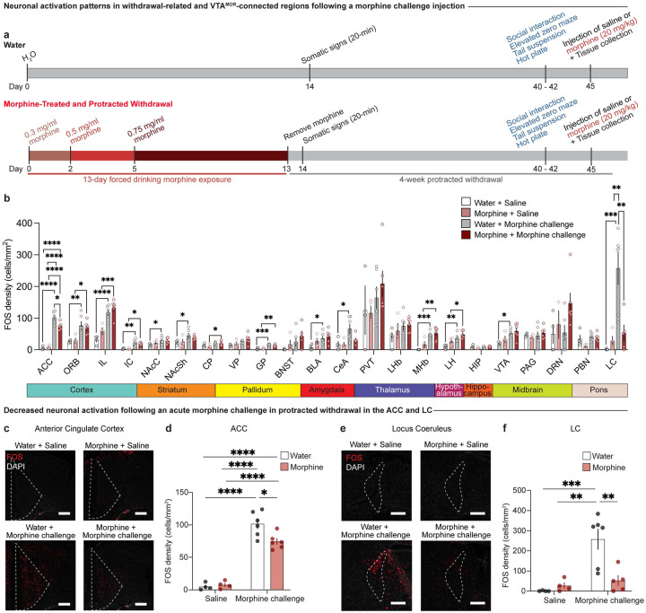
Figure 4: Protracted morphine withdrawal leads to decreased neuronal activation following acute morphine challenge in the ACC and LC.
(a) Experimental schematic of the 13-day chronic forced morphine drinking exposure with escalating concentrations, assessment of somatic withdrawal signs 24-hours following cessation of morphine administration, a 4-week protracted withdrawal period, behavioral battery assessment, and tissue collection 90 minutes following a morphine challenge injection. (b) Neuronal activation as represented by FOS density in opioid naïve and morphine-treated mice following a saline or morphine challenge injection (20 mg/kg, s.c.) across 22 regions connected to VTAMOR neurons or associated with withdrawal. Chronic morphine exposure results in decreased neuronal activation following an acute morphine challenge in protracted withdrawal in the (c and d) anterior cingulate cortex (two-way ANOVA + Bonferroni, treatment F1,16=4.312, p=0.0543; injection F1,16=190.3, p<0.0001; interaction F1,16=6.456, p=0.0218, scale, 200 μm) and (e and f) locus coeruleus (two-way ANOVA + Bonferroni’s multiple comparison, treatment F1,15=5.847, p=0.0288; injection F1,15=14.48, p=0.0017; interaction F1,15=9.757, p=0.0070, scale, 200 μm). Data are represented as mean ± SEM, *p<0.05, **p<0.01, ***p<0.001, ****p<0.0001.

