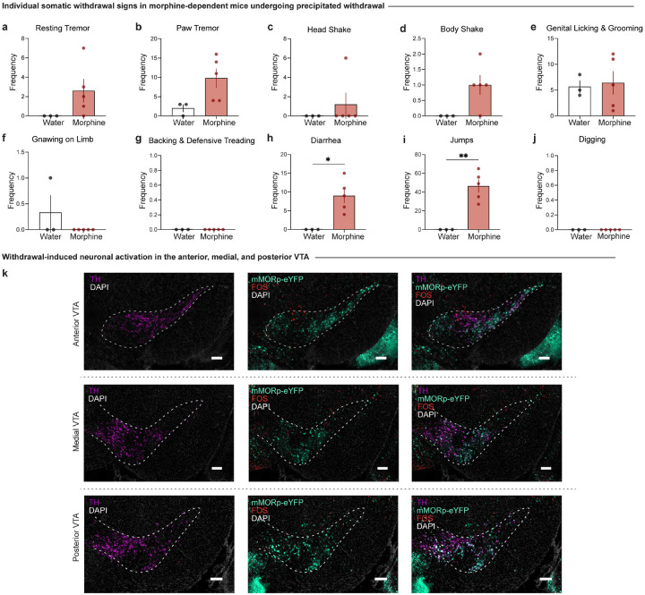
Extended Data Figure 2-1: Individual somatic withdrawal signs in morphine-dependent mice undergoing precipitated withdrawal and neuronal activation of MOR neurons in the anterior, medial, and posterior VTA.
Morphine-dependent mice undergoing precipitated withdrawal demonstrate increased frequencies of (h) diarrhea (unpaired t test, t=3.509, df=6, p=0.0127) and (i) jumps (unpaired t test, t=5.046, df=6, p=0.0023), while the frequencies of (a) resting tremors, (b) paw tremors, (c) head shakes, (d) body shakes, (e) genital licking and grooming, (f) gnawing on limb, and (g) backing and defensive treading bouts remained unchanged relative to opioid naïve mice. (k) Representative images of coronal sections showing tyrosine hydroxylase (TH) staining with mMORp-eYFP and FOS staining in the anterior, medial, and posterior VTA (scale, 200 μm). Data are represented as mean ± SEM, *p<0.05, **p<0.01. Scale, 200 μm.

