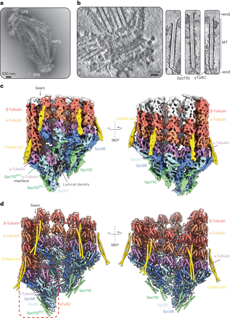
Fig. 1. Global architecture of the native budding yeast γTuRC capping MT minus ends.
a, A negative-stain electron micrograph of the S. cerevisiae SPBs forming a bipolar spindle, a typical structure observed following the enrichment procedure. Similar spindle structures were observed in more than ten independent SPB enrichment preparations. b, Left: a slice through a representative tomogram showing outer, central and inner plaques of the intact SPB, and a capped MT minus end (circled). Right: slices through denoised tomographic reconstructions of spindle MTs. MT-capping γTuRCs and Spc110 coiled coils connect the MTs to the SPB. The MT lattice and γTuRC caps are intact. c, A consensus cryo-ET reconstruction of the γTuRC capping the α/β-tubulin lattice after equalizing density thresholds using Occupy52. Scale bar as in b. d, A molecular model of the γTuRC bound to the MT minus end. Spokes are numbered as in Extended Data Fig. 2a, and a γTuSC unit is highlighted in a red box.

