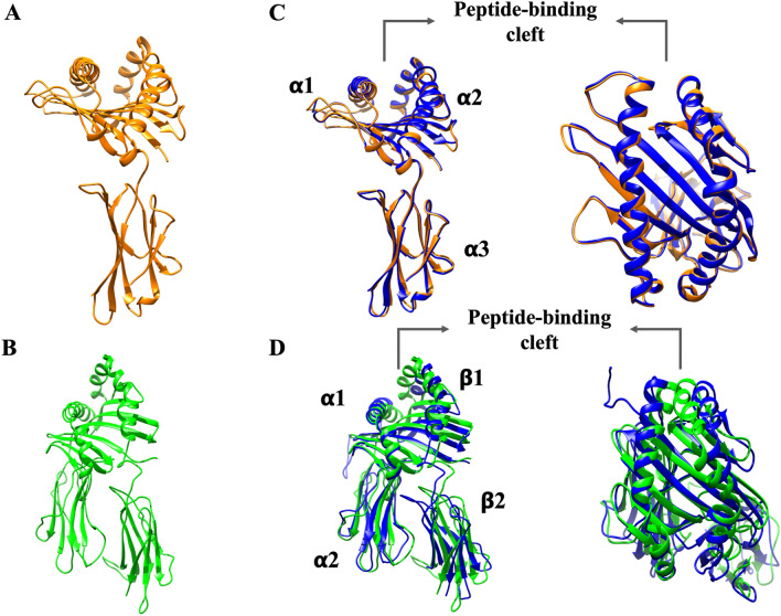
Figure 2.
Canine MHC molecule and TLR homology models obtained by MODELLER v. 10.0 and structural alignments with PDB template structures. (A) Canine MHC class I molecule homology model (orange). (B) Canine MHC class II molecule homology model (green). (C) Structural alignment of the template (PDB code: 5F1N) (blue) and canine MHC class I molecule homology model (orange). (D) Structural alignment of a template (PDB code: 4FQX) (blue) and canine MHC class I molecule homology model (green). Homology modeling was performed using MODELLER v. 10.0. The peptide-binding cleft was marked in both MHC molecules.

