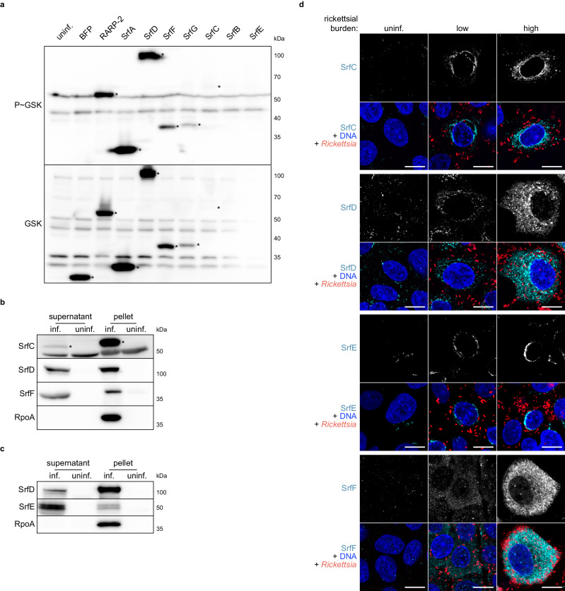
Fig. 3. Srfs are secreted by R. parkeri into the host cell during infection.
a Western blots for GSK-tagged constructs expressed by R. parkeri during infection of Vero cells. Whole-cell infected lysates were probed with antibodies against the GSK tag (bottom) or its phosphorylated form (P~GSK, top) to detect exposure to the host cytoplasm. BFP (non-secreted) and RARP-2 (secreted) were used as controls. SrfA–G are ordered by observed expression level. Asterisks indicate GSK-tagged protein bands. SrfB and SrfE (expected 37 and 50 kDa, respectively) were not detected. b Western blots for endogenous, untagged SrfC, SrfD, and SrfF during R. parkeri infection of A549 cells. Infected host cells were selectively lysed to separate supernatants containing the infected host cytoplasm from pellets containing intact bacteria. Asterisks indicate SrfC bands (apparent 55 kDa, but expected 48 kDa). SrfD and SrfF ran at the expected sizes (107 and 36 kDa, respectively). RpoA, lysis control. c Western blots for endogenous, untagged SrfD and SrfE during R. parkeri infection of A549 cells. Lysates were prepared as in b. SrfE ran at the expected size (48 kDa). RpoA, lysis control. d Images of Srfs (cyan) secreted by GFP-expressing R. parkeri (red) during infection of A549 cells (Hoechst, blue). Srfs were detected at both low and high rickettsial burdens. Scale bar, 10 μm. Uninfected host cells (uninf.) were included as controls for (a–d), and the results are representative of at least two independent experiments. Source data are provided as a Source Data file.

