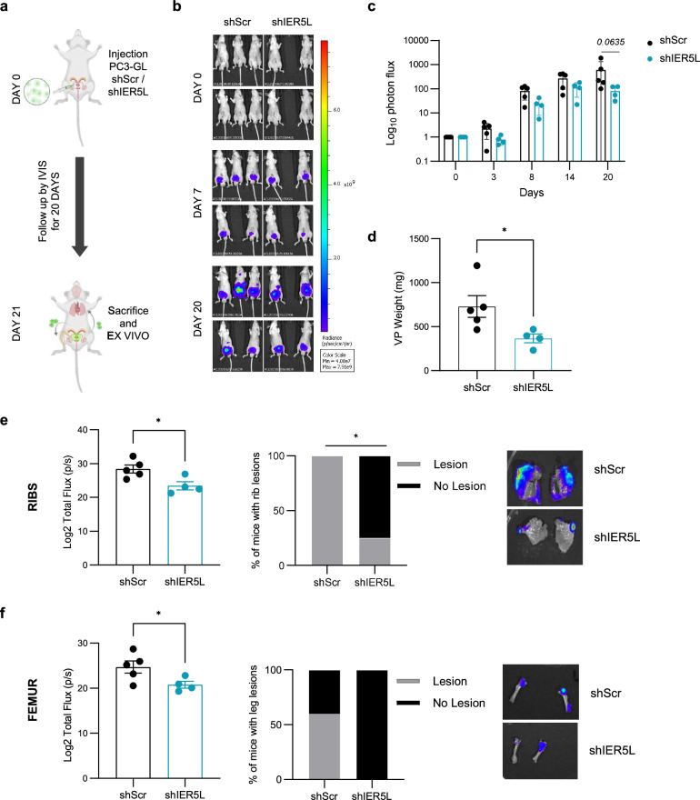
Fig. 4. Silencing of IER5L counteracts cell growth and metastatic dissemination of prostate cancer cells in vivo.
a Experimental design of the in vivo orthotopic assay. PC3 GFP-LUC (GL) cells transduced with shScramble (shScr) or sh2 IER5L (shIER5L) were injected into the ventral prostate lobe of nude mice and followed up for 20 days. IVIS relative flux data along the experimental process. Representative images (b) and the total photon flux normalized to time 0 (c) are represented. A multiple Mann-Whitney U-test was applied for statistical analysis. d Ex vivo tumor weight of the ventral lobes of prostates (VP) from the in vivo orthotopic assay. A two-tailed Mann-Whitney test was applied for statistical analysis. Ex vivo IVIS signal quantification of ribs (e) and femur (f) (left panels). Contingency analysis of metastatic lesions at those sites (middle panels) and a representative image (right panels) are shown. A one-tailed Mann–Whitney test and a Fisher exact t-test were used, respectively.

