
FIGURE 3.
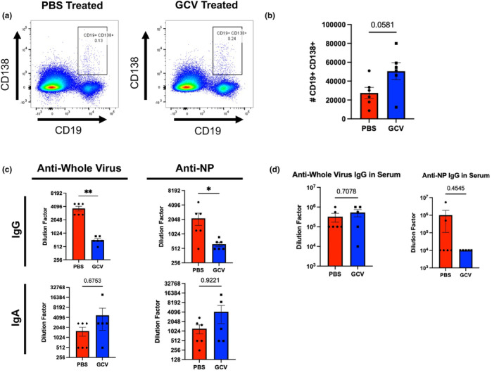
Targeting p16‐Expressing Cells Alters B‐cell Phenotypes and Antibody Secretion in the Lung. At 14 DPI, lung‐infiltrating B cells were identified as plasmablasts (CD19+ CD138+) and quantified (a,b). These were initially gated on CD4 and CD8 negative cells via a dump channel (as shown in Figure S4). Antibody titers were examined in bronchoalveolar lavage (BAL) to quantify IgA and IgG directed against either whole viral particles or flu nucleoprotein (NP) (c). IgG levels were quantified in the serum (d). All mice were infected using PR8. Data are presented as mean +/− standard error of the mean (SEM), and each symbol represents a single animal. Comparisons shown in B and top right panel of C were analyzed using Student's t‐test, all other comparisons were analyzed using the Mann–Whitney U‐test, all with a significance level of *p < 0.05; **p < 0.01. N = 5–6 per group (5 males in GCV group and 3 males in PBS group).