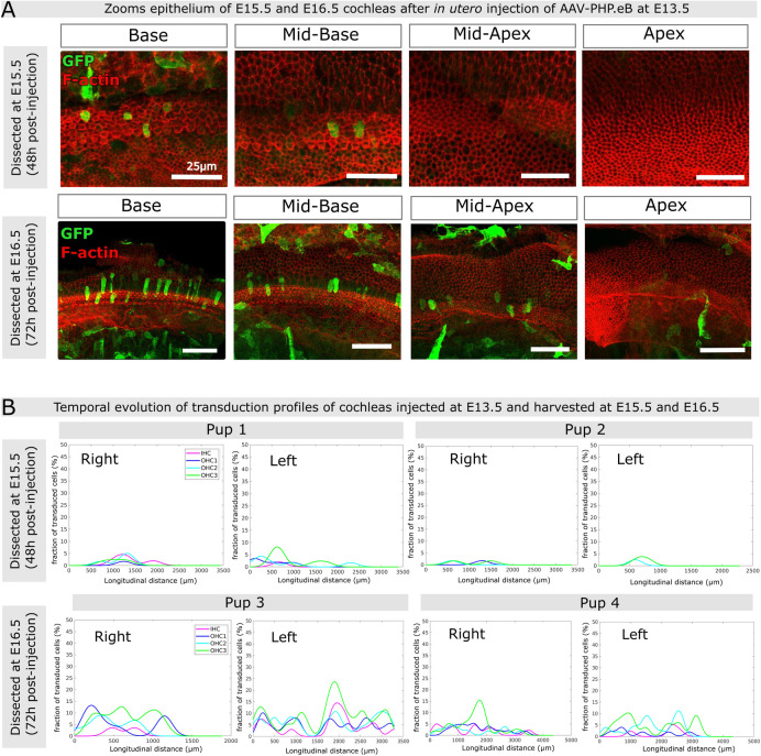
Fig 4. Inner ear transduction efficiency of AAV-PHP.eB administered at embryonic stages E15.5 and E16.5.
(A) Confocal views (maximum intensity projections) of the cochlear epithelium of mice subjected to unilateral injection of AAV-PHB.eB::GFP at E13.5, and imaged at E15.5 (upper panels) or at E16.5 (lower panels). Panels from left to right show different cochlear locations from base to apex. Staining for F-actin (red) and GFP (green). Scale bars 25 μm (upper panels) and 50 μm (lower panels). (B) Longitudinal transduction rate profiles observed in cochleas dissected at either E15.5 (upper panels) or E16.5 (lower panels) after unilateral injection of AAV-PHP.eB at E13.5. IHC, inner hair cells, OHC1, OHC2, and OHC3, first, second and third row of outer hair cells, respectively.

