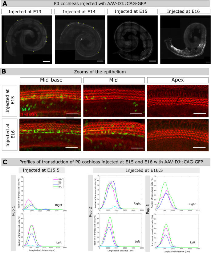
Fig 5. Inner ear transduction efficiency of AAV-DJ administered at embryonic stages.
(A) Whole-mount P0 cochleas injected with 1.2μl AAV-DJ at E13.5, E14.5, E15.5, E16.5. Scale bar 200 μm. (B) Close-ups of the mid-basal, mid, and apical regions of P0 cochleas injected either at E15.5 (upper panels), or at E16.5 (lower panels). Scale bar 25 μm. (C) Longitudinal transduction rate profiles measured in the right and left cochleas of three different pups injected at E15.5 (pup 1) or at E16.5 (pups 2 and 3). IphC, phalangeal cells, DC, Deiter cells, PC, pillar cells, IBC, inner border cells.

