Approximately 10% of metastases from all primary neoplasms involve the skin, but for malignant melanoma the figure is 44%.1 In some cases of melanoma this is the presenting feature, either because the primary lesion has regressed completely or because it has been unnoticed or ignored by the patient. Occasionally the melanoma has originated at an extracutaneous site such as the retina or the anal canal.
Metastases from cutaneous melanoma normally present as flesh coloured papules or nodules in the skin. Only about a third are pigmented or ulcerated. We report a case in which cutaneous metastases from a melanoma imitated herpes zoster. This presentation is known as zosteriform metastasis; it also occurs with other neoplasms.
Case report
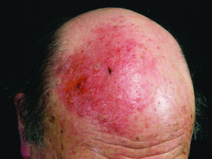
A 73 year old white man presented with a three week history of painful, pruritic vesicles on a background of erythema on the right frontal area of the scalp (figure). The lesion had not responded to self prescribed topical antibiotics and antiseptics. The patient had grown up in South Africa and had a history of excessive exposure to the sun. He had previously developed three basal cell carcinomas and had numerous actinic keratoses. Five years earlier a malignant melanoma, Breslow thickness 1.25 mm, had been excised from his right shoulder. At that time there had been no evidence of metastatis. He had been followed up regularly and there had been no evidence of recurrence. He also had a five year history of stage Ib mycosis fungoides.
Clinical examination showed that the lesion lay within the area supplied by the ophthalmic branch of the right trigeminal nerve. A provisional diagnosis of herpes zoster (shingles) was made, and he was treated with 800 mg of oral aciclovir five times a day. There was no history of herpes zoster at the same site or elsewhere.
He was reviewed seven days after starting aciclovir. The lesions had extended slightly but otherwise remained unchanged. At that point a diagnosis of plaque stage mycosis fungoides was considered, and the scalp lesion was biopsied. Histological examination showed that the dermis was heavily infiltrated by non-pigmented malignant cells, which were epithelioid in appearance and forming nests; immunostaining showed that these cells were metastatic melanoma.
The area was too extensive to excise. After discussion with the patient the lesion was treated with electron beam radiotherapy (40 Gy in 15 fractions). The response was dramatic and within a few weeks the lesion regressed almost completely except for some residual macular erythema.
Discussion
Many different malignant tumours can metastasise to the skin but the commonest primary sources are tumours of breast, stomach, lung, and uterus. These lesions usually present as firm papules or nodules, both of which may ulcerate; occasionally they are inflammatory, sclerotic, bullous, or vesicular. Zosteriform metastasis is less well known; it may arise from adenocarcinoma of the lung,2,3 carcinoma of the prostate,4,5 Kaposi's sarcoma,6,7 transitional cell carcinoma of the bladder,8 or malignant melanoma.9,10 Zosteriform metastases are usually painful, tender, or pruritic and consist of vesicles on a background of erythema, imitating the appearance of shingles. They are commonly confined to a single unilateral dermatome, adding to the potential for misdiagnosis. Our patient presented with zosteriform metastases which we initially misdiagnosed as herpes zoster.
It is obviously important to recognise metastatic disease. Firstly, it may indicate an occult malignancy and call for a vigorous search for the primary lesion, which may be internal; if the primary lesion is cutaneous it may have been unnoticed or ignored by the patient. Secondly, metastatic disease may represent progression of a known malignancy, requiring a change in management.
The mechanism for the zosteriform appearance of metastatic disease is not known. It has been postulated that recent herpes zoster might induce infiltration of malignant cells in a Koebner-like phenomenon.6,11 Our patient had no history of herpes zoster or any other skin lesion in this area. Perineural lymphatic spread of malignant cells has been suggested as a mechanism2,4,5,12; invasion of the dorsal root ganglia with subsequent peripheral spread is another hypothesis.3,8,12 Our case also highlights the value of histological examination in the management of skin lesions that do not respond to treatment.
Figure.
Zosteriform metastasis from malignant melanoma
Footnotes
Cutaneous metastases from primary tumours, including malignant melanoma, can imitate herpes zoster (shingles)
Funding: None.
Competing interests: None declared.
References
- 1.Lookingbill DP, Spangler N, Helm KF. Cutaneous metastases in patients with metastatic carcinoma: a retrospective study of 4020 patients. J Am Acad Dermatol. 1993;29:228–236. doi: 10.1016/0190-9622(93)70173-q. [DOI] [PubMed] [Google Scholar]
- 2.Hodge SJ, Mackel S, Owen LG. Zosteriform inflammatory metastatic carcinoma. Int J Dermatol. 1979;18:142–145. doi: 10.1111/j.1365-4362.1979.tb04492.x. [DOI] [PubMed] [Google Scholar]
- 3.Matarasso SL, Rosen T. Zosteriform metastases:case presentation and review of the literature. J Dermatol Surg Oncol. 1988;14:774–778. doi: 10.1111/j.1524-4725.1988.tb01162.x. [DOI] [PubMed] [Google Scholar]
- 4.Braverman IM. Cancer. Skin metastases and related disorders. In: Braverman IM, editor. Skin signs of systemic disease. 2nd ed. Philadelphia: Saunders; 1981. pp. 1–14. [Google Scholar]
- 5.Bluefarb SM, Wallk S, Gecht M. Carcinoma of the prostate with zosteriform cutaneous lesions. Arch Dermatol. 1957;76:402–406. doi: 10.1001/archderm.1957.01550220010002. [DOI] [PubMed] [Google Scholar]
- 6.Niedt GW, Prioleau PG. Kaposi's sarcoma occurring in a dermatome previously involved by herpes zoster. J Am Acad Dermatol. 1988;18:448–451. doi: 10.1016/s0190-9622(88)70068-7. [DOI] [PubMed] [Google Scholar]
- 7.Roth JS, Grossman ME. Linear Kaposi's sarcoma in HIV disease. J Am Acad Dermatol. 1993;29:488. doi: 10.1016/s0190-9622(08)82003-8. [DOI] [PubMed] [Google Scholar]
- 8.Jaworsky C, Bergfield WF. Metastatic transitional cell carcinoma mimicking zoster sine herpete. Arch Dermatol. 1986;122:1357–1358. [PubMed] [Google Scholar]
- 9.Itin PH, Lautenschlager S, Buechner SA. Zosteriform metastases in melanoma. J Am Acad Dermatol. 1995;32:854–857. doi: 10.1016/0190-9622(95)91546-x. [DOI] [PubMed] [Google Scholar]
- 10.North S, Mackey JR, Jensen J. Recurrent malignant melanoma presenting with zosteriform metastases. Cutis. 1998;62:143–146. [PubMed] [Google Scholar]
- 11.Aloi FG, Appino A, Puiatti P. Lymphoplasmocytoid lymphoma arising in herpes zoster scars. J Am Acad Dermatol. 1990;22:130–131. doi: 10.1016/s0190-9622(08)80015-1. [DOI] [PubMed] [Google Scholar]
- 12.Manteaux A, Cohen PR, Rapini RP. Zosteriform and epidermotropic metastasis. Report of two cases. J Dermatol Surg Oncol. 1992;18:97–100. doi: 10.1111/j.1524-4725.1992.tb02440.x. [DOI] [PubMed] [Google Scholar]


